NUCLEOTYPE
Electrocradiogram Findings in Hyperkalemia
January 20, 2020
Potassium plays an essential role in the electrical conduction through the heart. When serum potassium levels are higher than 5.5 mEq/L, there becomes an increased risk for lethal cardiac dysrhythmias and cardiac arrest.
Hyperkalemia is defined as a serum potassium level greater than 5.5 mEq/L. After this, you’ll start to see a step-wise progression of cardiac conduction abnormalities. However, it is important to note that the rate at which the serum potassium level rises is strongly related to the severity of the electrocardiogram (ECG) changes. For example, patients who have chronically elevated serum potassium levels may have a relatively normal ECG.
Generally, you’ll want to be aware of some arbitrary levels after which you’ll start to see specific ECG changes:
- Mild - serum potassium > 5.5 mEq/L
- Moderate - serum potassium > 6.5 mEq/L
- Severe - serum potassium > 7.0 mEq/L
- Arrest - serum potassium > 9.0 mEq/L
Serum Potassium level > 5.5 mEq/L
Repolarization abnormalities
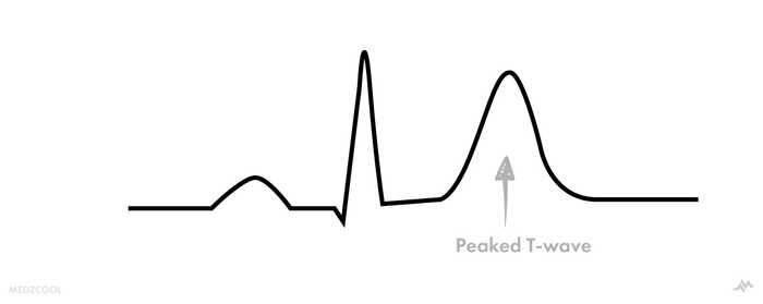
Remember repolarization of the ventricles on an EKG strip is represented as the T-wave. Therefore, repolarization abnormalities related to hyperkalemia affect the T-waves, specifically causing them to be “peaked”.
Peaked T-waves are usually the first findings found on the ECG in the setting of hyperkalemia.
Serum Potassium level > 6.5 mEq/L
At serum potassium levels greater than 6.5 mEq/L, the cardiac rhythm strip’s intervals and waves continue to lengthen, flatten and heart rate slows.
Wide and Flat p-waves
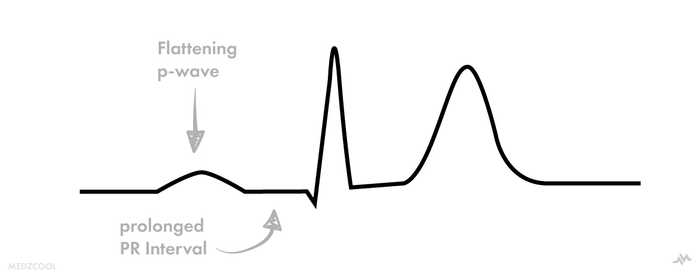
At this level, p-waves start to widen and flatter. Then, intervals begin to lengthen, starting with the PR segment.
Remember that first degree heart blocks are defined as PR intervals greater than 200 ms, and in some cases of mild hyperkalemia, you may start noticing various atrioventricular blocks.
Serum Potassium level > 7.0 - 9.0 mEq/L
At levels greater than 7.0 mEq/L, you continue to see the lengthening of intervals, including the complete flattening of the p-wave.
Widening QRS Complexes
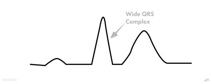
QRS widening occurs at levels > 7.0 mEq/L, in addition to slowed heart rates. This may provoke the development of ventricular escape rhythms in the setting of heart blocks, bundle branch blocks, fascicular blocks, etc.
Sinusoidal Wave Pattern
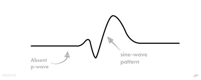
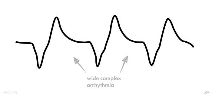
As the potassium level starts to get closer to 9.0 mEq/L, the ECG rhythm strip starts to develop a sine-wave morphology. This is potentially a terminal rhythm suggesting possible deterioration into a life-threatening arrhythmia.
Think of the sinusoidal wave pattern as a potential warning sign for cardiac arrest (PEA, asystole, ventricular fibrillation, etc)
Serum Potassium level > 9.0 mEq/L
Dysrhythmias and Cardiac Arrest
At these levels, you’ll start to see patients in cardiac arrest. The sine-wave morphology will progress into a wide-complex rhythm and then pulseless arrest, asystole, and in some cases, ventricular fibrillation.
Remember, these levels are not specific cut-off points and that patients may present with ECG changes that do not necessarily correlate to the points mentioned above. So it’s important not to use these points as thresholds for determining risk or treatment.
