NUCLEOTYPE
First Versus Second Degree Heart Block
June 05, 2020
In normal sinus rhythm, there is a P-wave that precedes each QRS complex and in between these two, is a fixed PR interval ranging from 120 to 200 milliseconds. In atrioventricular (AV) blocks, there is a delay in the conduction of electrical impulses from atria to the ventricles which can be the result of an anatomical or functional abnormality in the conduction system of the heart.
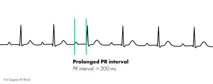
In first degree AV blocks, there is a delay in electrical conduction from atria to ventricles. This manifests on an electrocardiogram (ECG), as a PR interval greater than 200 milliseconds.
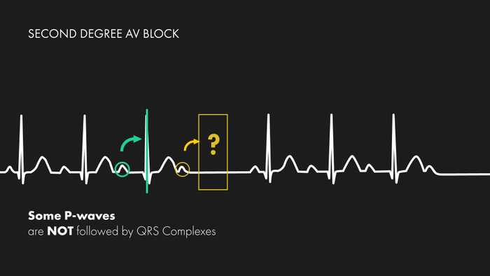
In second degree AV blocks, there is intermittent conduction of atrial impulses to the ventricles resulting in some P-waves, not being followed by QRS complexes. This relationship of P:QRS often occurs with regularity, such as in ratios of 2:1, 3:2, 4:3, etc. Second degree AV blocks can also be further divided into Mobitz type 1 (Wenckebach) or Mobitz type 2, which can be differentiated by examining the PR intervals.
What’s the difference?
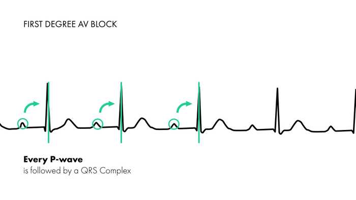
The difference between first and second degree AV blocks is in the relationship between P-wave:QRS-complex. Regardless of the length or variations in the PR interval, if some P-waves are not followed by QRS-complexes, (if there are “missing” QRS complexes), then the rhythm must be a second-degree block or higher. Remember, in first-degree heart blocks, every P-wave is followed by a QRS complex.
Notice in the image above, every P-wave is followed by a QRS. The rhythm may appear normal sinus at first, however on further inspection of the intervals, you’ll find that the PR interval is longer than 200 milliseconds.
In second-degree heart blocks, as seen in the photo above, if you follow each P-wave, you’ll notice that there are times when a P-wave is not followed by a QRS complex. There is a pause in cardiac activity until normal conduction appears to restart again with P-QRS complexes. Following this rhythm further will also reveal that this relationship of P:QRS occurs at regular intervals.
Further evaluation of the PR interval in cases of second-degree heart blocks can further distinguish whether the rhythm is a Mobitz type 1 or Mobitz type 2 second degree AV block, but is discussed in another article (Type 1 versus Type 2 Second Degree Heart Block).


