NUCLEOTYPE
The Cardiac Cycle (P-QRS-T)
November 01, 2021

The waves and bumps you see on the EKG represents electrical depolarization moving through the heart, which can correlate to the different stages of the cardiac cycle and to cardiac contraction.
Normally, depolarization starts at the Sino-Atrial Node (SA Node) and propagates to adjacent cardiac myocytes until electricity has made its way throughout the entirety of the heart.
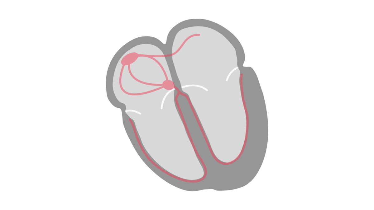
The Pacemaker

The SA Node is the heart’s dominant pacemaker. It is located in the upper-posterior wall of the right atrium initiating a depolarization wave at regular intervals.
Atrial Depolarization

Each depolarization wave of positive charges proceeds outward from the SA Node and stimulates both atria to contract. Atrial depolarization is recorded and visualized on the EKG as a P wave.
AV Node and the PR Interval
Depolarization from the atrial conduction system eventually propagates to the next site in the heart which carries specialized cells used in electrical conduction - the AV node.

When depolarization reaches the AV node from the atria, electrical conduction is slowed, before it propagates down to the ventricles. This slowing of electrical conduction is represented on the EKG as the PR interval, which is measured from the beginning of the P wave to the start of the QRS complex. It reflects the conduction through the AV node. This is also the time when the atrial myocytes begin to repolarize.

The QRS Complex
This pause in electrical activity, allows enough time for blood to empty from the atria into the ventricles, just before ventricular depolarization and contraction.

After the Bundle of His, the Purkinje fibers bifurcate, leading depolarization to continue down the Left and Right Bundle Branches of the ventricles and eventually to the ventricular myocardium. Depolarization of the ventricular myocardium records as a QRS complex, which leads to ventricular contraction.

The Q wave is the first downward deflection of the QRS complex. It is not always present on an EKG, in fact in most cases, you may not see an initial downward deflection and so no Q wave. When a Q wave is present, is it followed by an upward deflection, the R wave. Any downward deflection after an upward wave is an S wave.
Ventricular Repolarization

Following the QRS complex, is a segment of horizontal baseline known as the ST segment. The ST segment represents the initial phase of ventricular repolarization. Remember that repolarization occurs so that the ventricular myocytes can recover their interior, resting negative charge so they can be depolarized again during the next cardiac cycle.
Repolarization of the ventricular myocytes begins immediately after the QRS and persists until the end of the T wave, which is upward wave following the ST segment.

In relation to ventricular contraction, remember that it begins at the start of the QRS complex and persists until the end of the T wave. This means that ventricular contraction spans depolarization and repolarization of the ventricles. This can be visualized on the EKG as the QT interval.

The QT interval represents the duration of ventricular systole (contraction) and is measured from the beginning of the QRS complex until the end of the T wave.
Summary
A cardiac cycle is represented by the P wave, the QRS complex, the T wave, and the baseline that follows until another P wave appears. This cycle is repeated continuously in coordination with SA node depolarization.
Atrial depolarization (and contraction) is represented by the P wave. Ventricular depolarization (and contraction) is represented by the QRS complex. During this time, the atria are undergoing and completing repolarization, but you don’t see atrial repolarization represented on the EKG because it occurs at approximately the same time as the QRS complex. As a result atrial repolarization is “buried” within the QRS complex.
Unlike atrial repolarization, ventricular repolarization is clearly visualized on the EKG as the ST segment, ending with the T wave.
This P-QRS-T represents one complete cardiac cycle.